
Best Time for 3D Ultrasound in Pregnancy | Timing Guide
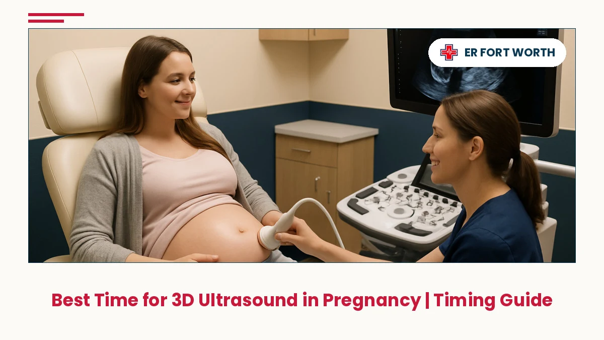
Understanding when to get 3D ultrasound in pregnancy is crucial for capturing clear images of your developing baby while ensuring the safest imaging windows. This guide walks you through trimester timing, fetal development milestones, and what to expect at each stage.
In This Article
- When to Get 3D Ultrasound in Pregnancy: Basic Timing
- Second Trimester: The Optimal Imaging Window
- First Trimester 3D Ultrasound Timing & What to Expect
- Third Trimester Imaging: Timing, Positioning & Clarity
- Fetal Development Stages & Imaging Windows
- Scheduling Tips for the Best 3D Ultrasound Results
- When to Seek Urgent Ultrasound Evaluation in Fort Worth
- Frequently Asked Questions
⚡ Key Takeaways
- The second trimester (18–24 weeks) offers the clearest 3D ultrasound images due to adequate amniotic fluid and fetal positioning.
- First trimester 3D scans (11–14 weeks) are possible but carry higher risk of motion artifact and less detailed facial features.
- Third trimester imaging (28+ weeks) is harder to visualize due to crowding and bone ossification, making fetal positioning critical.
- Optimal fetal development windows exist for each trimester—understanding these windows helps your provider plan the best imaging timing.
- Scheduling 3D ultrasounds during recommended milestones allows you to capture the most emotionally meaningful and clinically useful images.
When to Get 3D Ultrasound in Pregnancy: Basic Timing
When to get 3D ultrasound in pregnancy depends on what you hope to capture and how well your baby is positioned. While 2D ultrasounds are standard throughout pregnancy for medical monitoring, 3D ultrasounds are primarily elective and provide enhanced visualization of facial features, limb positioning, and structural details. The timing directly affects image quality—earlier pregnancies may produce cloudier images, while later pregnancies face crowding and reduced fluid volume that obscure detail.
Most obstetricians and maternal-fetal specialists recommend scheduling elective 3D ultrasounds during the second trimester, specifically between 18 and 24 weeks of gestation. This window balances fetal size, amniotic fluid volume, and bone development to yield the clearest three-dimensional renderings. However, some providers offer first trimester 3D imaging as early as 11–14 weeks, and third trimester scans are possible, though with specific considerations around fetal positioning and imaging clarity.
If you're experiencing pregnancy complications or have medical questions about ultrasound timing in the Fort Worth, Keller, Saginaw, or North Richland Hills area, ER Of Fort Worth provides 24/7 access to care for urgent maternal health concerns. Always discuss ultrasound timing and necessity with your obstetrician, as they can advise based on your individual pregnancy and medical history.
Second Trimester: The Optimal Imaging Window
The second trimester—specifically weeks 18 through 24—represents the gold standard for 3D ultrasound imaging. During this window, your baby has grown large enough to produce detailed facial and limb images, yet remains in a relatively spacious amniotic sac with ample fluid surrounding them. The fetal skeleton is still partially cartilaginous rather than fully ossified (hardened to bone), which allows ultrasound waves to penetrate and visualize soft tissue contours with exceptional clarity.
Around 20 weeks, many families schedule their anatomy scan, which is the medically indicated standard ultrasound to assess fetal development, organ formation, and screen for structural anomalies. Some providers bundle a 3D component into this appointment or recommend a separate 3D session shortly after. At 22–24 weeks, fetal features are well-developed enough for stunning three-dimensional renderings of facial structure, hand positioning, and profile views. The amniotic fluid is still plentiful, minimizing shadowing artifacts that can cloud images later in pregnancy.
Your baby's positioning during this trimester also tends to be more variable, meaning they may shift naturally to favorable angles for imaging. If you're in Fort Worth, Grapevine, Haslet, or the surrounding service areas and want to discuss elective ultrasound timing as part of your prenatal care, your obstetrical provider can coordinate the best appointment slot. Remember that while elective 3D ultrasounds are generally considered safe based on established medical consensus, they should be performed by qualified sonographers or physicians using appropriate ultrasound settings.
First Trimester 3D Ultrasound Timing & What to Expect
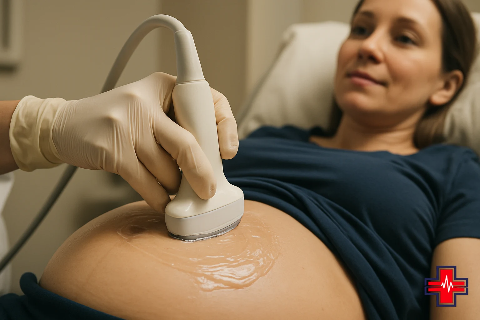
First trimester 3D ultrasound imaging is possible between 11 and 14 weeks of gestation and has become increasingly popular for early prenatal bonding and assessment. This window captures your baby during the nuchal translucency (NT) scan period, when certain markers can be measured to assess risk for chromosomal abnormalities. However, first trimester 3D imaging presents distinct challenges compared to second trimester timing.
At 11–14 weeks, your baby is significantly smaller—crown-to-rump length is roughly 2 to 3 inches. While detailed facial features are beginning to form, the fetal face is still compact and may not render with the same dimensional clarity as in the second trimester. Amniotic fluid volume is also more limited, which can reduce the imaging window and create more shadowing. Additionally, first trimester fetuses are often in motion, leading to motion artifact that blurs the 3D reconstruction. Many sonographers find that obtaining quality 3D images during this period requires extended scanning time and patient positioning adjustments.
If you're considering first trimester 3D ultrasound, discuss realistic expectations with your provider. Some pregnancies will yield beautiful early images; others may require a follow-up second trimester scan for optimal results. If you have concerns about pregnancy development or need urgent ultrasound evaluation in Fort Worth, Keller, Watauga, or nearby areas, contact your obstetrical clinic or ER Of Fort Worth for immediate guidance.
Third Trimester Imaging: Timing, Positioning & Clarity
Third trimester 3D ultrasound imaging—from 28 weeks onward—is technically feasible but presents significant challenges that many families and providers prefer to avoid. By the third trimester, your baby has grown substantially larger, consuming more of the amniotic sac and reducing the fluid volume that surrounds them. This crowding naturally limits the acoustic windows available for imaging and increases the likelihood of fetal shadowing, where bony structures or positioning block clear visualization of facial features and body contours.
Additionally, fetal bone ossification (hardening) accelerates in the third trimester. While this is a normal and healthy developmental process, increased bone density reduces ultrasound penetration, making soft tissue detail harder to render in three dimensions. Many third trimester fetuses also assume head-down (vertex) positioning in preparation for delivery, which can make capturing anterior facial views or full-body 3D images challenging. Posterior or lateral positioning during this period often yields inadequate images.
Third trimester 3D ultrasounds are sometimes requested to assess suspected structural concerns identified earlier in pregnancy, but elective third trimester imaging for bonding or keepsake purposes is generally less satisfying than second trimester scans. If you do pursue a third trimester 3D ultrasound, sonographers may ask you to change positions, walk, or even return for a second appointment if the first session doesn't yield adequate images. For any pregnancy-related concerns in Fort Worth, Southlake, Roanoke, or surrounding communities, your obstetrician is the best resource for deciding whether additional imaging is necessary and when to schedule it.

Fetal Development Stages & Imaging Windows
Understanding fetal development stages helps explain why ultrasound imaging windows matter. During the first trimester (weeks 0–13), organogenesis—the formation of major organs—is the primary developmental task. The embryo grows from a few millimeters to about 3 inches and transitions from embryonic to fetal form around 8–9 weeks. By 11–14 weeks, facial features, limbs, and major organs are recognizable, but detail remains limited due to small size.
The second trimester (weeks 14–27) is characterized by rapid growth and maturation. Facial features become increasingly distinct and proportionate. Limbs lengthen and fine motor movements develop. By mid-second trimester (18–20 weeks), fetal proportions resemble a newborn more than an embryo, and the anatomy scan can visualize all major organ systems. This is also when fetal body fat begins accumulating, which softens facial contours and produces more lifelike 3D renderings. Between 20 and 24 weeks, the fetal face reaches an aesthetic peak for imaging—detailed enough for stunning facial features yet still maintaining rounded proportions that look "cute" to parents viewing keepsake images.
The third trimester (weeks 28–40) is dominated by growth, accumulation of body fat and glycogen, lung maturation, and positioning for delivery. By 30+ weeks, the fetus may occupy 90% of the amniotic sac, leaving minimal imaging space. Bone ossification accelerates, and skin texture becomes more opaque. These changes collectively explain why third trimester 3D ultrasounds often yield less satisfying results than earlier scans. For pregnancy monitoring and urgent questions in Fort Worth, Flower Mound, Bedford, and neighboring areas, ER Of Fort Worth is available 24/7 to address maternal concerns.
Scheduling Tips for the Best 3D Ultrasound Results
Timing is only one factor in obtaining excellent 3D ultrasound images. Several practical steps can optimize your appointment. First, schedule your 3D ultrasound during the second trimester sweet spot—ideally 20–23 weeks. This gives your baby time to develop distinct features while remaining in a spacious, fluid-filled environment. Avoid scheduling too early in the second trimester (before 18 weeks), as facial definition may still be developing.
Second, maintain good hydration in the days and hours before your appointment. Adequate amniotic fluid improves acoustic quality and gives your baby room to move and position favorably. Some providers recommend eating a light snack beforehand, as fetal movement often increases after maternal glucose intake, improving the chances of capturing active, well-positioned images.
Third, wear comfortable clothing that allows easy access to your abdomen. Most 3D ultrasounds take longer than standard 2D scans—often 20–30 minutes or more—because sonographers carefully angle the transducer to capture multiple planes of imaging data that the machine then reconstructs into 3D images. Patience and comfortable positioning on your part facilitate this process.
Fourth, discuss your preferences with your sonographer or physician before the scan begins. If you have specific images in mind (profile view, hand near face, full-body pose), mention this so they can prioritize capturing those angles. Finally, arrange for a support person—partner, family member, or friend—to accompany you, as they can see the images in real-time and the experience is often more meaningful when shared. For any urgent maternal health concerns in the Fort Worth, Keller, or Alliance areas, your obstetrician or ER Of Fort Worth can provide immediate support.
When to Seek Urgent Ultrasound Evaluation in Fort Worth
While most 3D ultrasounds are elective and can be scheduled at your convenience, certain pregnancy symptoms warrant urgent ultrasound evaluation. If you experience vaginal bleeding, severe abdominal pain, sudden onset of dizziness or shortness of breath, or loss of fetal movement (particularly after 20 weeks when you should feel regular movement), contact your obstetrician or seek emergency care immediately. If you are experiencing a life-threatening emergency, call 911 or go to the nearest emergency room immediately.
ER Of Fort Worth, located at 4561 Heritage Trace Parkway, Suite 117, is available 24/7 to evaluate urgent pregnancy-related concerns. If you cannot reach your obstetrician during evenings, weekends, or holidays, our emergency team can perform bedside ultrasound assessment, check your vital signs, and determine whether your symptoms require intervention or specialist consultation. We serve families from Fort Worth, Keller, Saginaw, Haslet, Watauga, Roanoke, North Richland Hills, Haltom City, Bedford, Southlake, Grapevine, Flower Mound, and Alliance.
Urgent ultrasound indications include threatened miscarriage (cramping with bleeding in first or early second trimester), suspected ectopic pregnancy (positive pregnancy test with pain and no intrauterine pregnancy visible), polyhydramnios or oligohydramnios concerns (too much or too little fluid), decreased fetal movement, and maternal trauma or injury during pregnancy. Our medical team can provide initial assessment and coordinate care with your obstetrical provider. Call 817-945-4200 or visit our Fort Worth emergency room location for immediate evaluation.
Frequently Asked Questions
What is the best week in pregnancy for 3D ultrasound imaging?
Weeks 20–24 (late second trimester) offer the optimal balance of fetal size, facial detail, amniotic fluid volume, and bone development. This window produces the clearest, most lifelike 3D images. Weeks 18–19 are also acceptable, though facial features may be slightly less defined; weeks 25+ are generally less ideal due to crowding and reduced fluid.
Can I get a 3D ultrasound in the first trimester?
Yes, 3D ultrasound is possible between 11–14 weeks during the NT scan window. However, image quality is typically less detailed than second trimester scans due to smaller fetal size, limited amniotic fluid, and increased fetal movement. Many families choose to wait until the second trimester for better results, though some first trimester scans yield beautiful images if your baby cooperates.
Why are third trimester 3D ultrasounds often unclear?
By 28+ weeks, your baby occupies most of the amniotic sac, reducing imaging space. Bone ossification increases, blocking ultrasound penetration. Fetal positioning (often head-down) makes anterior facial views difficult. These factors combine to reduce clarity compared to second trimester imaging.
Is it safe to have multiple 3D ultrasounds during pregnancy?
Yes, 3D ultrasound is generally considered safe when performed by qualified professionals using appropriate settings and without excessive scanning duration. Discuss the frequency and necessity of additional ultrasounds with your obstetrician, as they can advise based on your pregnancy and medical history.
What should I do if I have pregnancy symptoms and need urgent ultrasound?
Contact your obstetrician immediately. If it is after hours, weekends, or holidays and symptoms are urgent (bleeding, severe pain, decreased fetal movement, shortness of breath), go to your nearest emergency room or call 911. ER Of Fort Worth, available 24/7 at 817-945-4200, can perform urgent bedside ultrasound evaluation and coordinate with your obstetrical team.
Can I request a specific baby pose or angle during my 3D ultrasound?
Yes, discuss your preferences with your sonographer before the scan. However, the baby's position is ultimately beyond your control. If your baby is positioned unfavorably on the first attempt, the sonographer may ask you to move, walk, or return for a follow-up appointment to capture the desired images.