
3D vs 2D Ultrasound: What's the Difference?
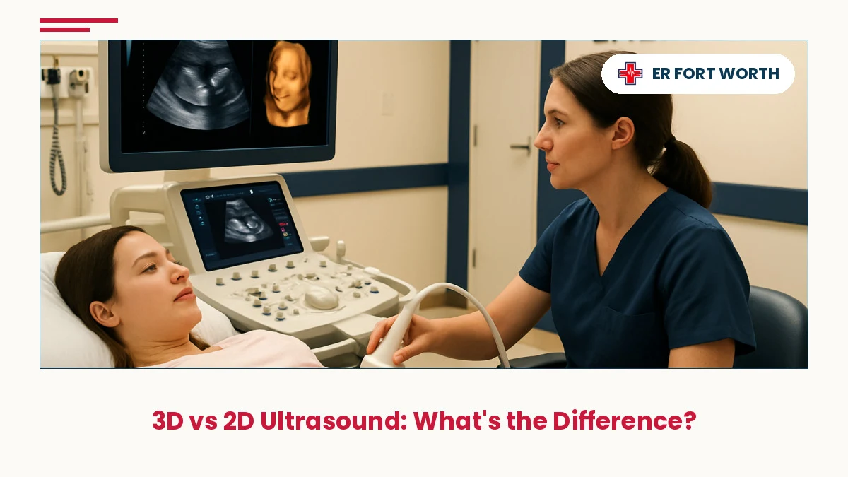
Ultrasound technology has transformed prenatal care, but expectant parents in Fort Worth and surrounding areas often wonder about the differences between 2D and 3D imaging. Understanding how these modalities compare helps you make informed decisions about your pregnancy monitoring.
In This Article
⚡ Key Takeaways
- 2D ultrasound uses flat, cross-sectional images and remains the clinical gold standard for diagnosing fetal anomalies and monitoring development.
- 3D ultrasound creates volumetric reconstructions that allow viewing from multiple angles, offering enhanced visualization for certain anatomical features.
- Both 2D and 3D ultrasound are safe when performed according to established medical guidelines, using appropriate thermal and mechanical indices.
- Diagnostic accuracy depends on operator skill, equipment quality, and clinical indication—not simply on choosing 3D over 2D imaging.
- Freestanding emergency rooms like ER Of Fort Worth provide rapid ultrasound assessment for urgent pregnancy concerns including bleeding, pain, and viability questions.
What Is 2D Ultrasound Imaging?
Two-dimensional (2D) ultrasound is the conventional imaging modality used in prenatal care for decades. It works by transmitting high-frequency sound waves into the body and recording the echoes that bounce back from tissues, organs, and the developing fetus. These echoes are converted into grayscale images displayed in real time on a monitor, showing flat, cross-sectional "slices" of anatomy.
In pregnancy, 2D ultrasound allows clinicians to measure fetal size, evaluate organ development, assess amniotic fluid volume, and detect structural abnormalities. The images are acquired in multiple planes—transverse, sagittal, and coronal—to build a comprehensive understanding of fetal anatomy. Because 2D ultrasound is performed in real time, it captures fetal movement and function, making it ideal for evaluating blood flow patterns and cardiac function. This modality has an established evidence base spanning decades and remains the clinical reference standard for routine prenatal screening worldwide.
The 2D approach provides immediate, dynamic information that supports clinical decision-making. Trained sonographers and physicians can perform targeted assessments based on clinical questions, adjusting imaging planes on the fly to answer specific diagnostic concerns. This flexibility and real-time nature make 2D ultrasound indispensable in emergency settings, where rapid assessment of fetal well-being or maternal complications is critical.
What Is 3D Ultrasound Imaging?
Three-dimensional (3D) ultrasound represents an evolution in imaging technology, building on the same basic principle as 2D but with added dimensionality. Instead of acquiring single planes of sound, 3D ultrasound systems rapidly capture multiple 2D images in quick succession across a volume of tissue. Specialized software then reconstructs these individual slices into a three-dimensional volumetric dataset that can be rotated, tilted, and viewed from any angle on the workstation.
The key advantage of 3D ultrasound is that once the volume has been acquired, the operator can manipulate and reorient it without sending the transducer back to the patient. This "post-processing" capability allows for detailed surface renderings—sometimes called 4D when motion is added in real time—that create photorealistic images of fetal features such as facial anatomy, hand position, and body contours. Some patients and families find these detailed images emotionally meaningful and enjoy the enhanced visualization of their developing baby.
From a clinical perspective, 3D ultrasound adds value in specific scenarios: evaluating cleft palate, assessing complex cardiac lesions, or providing orthogonal views of anatomical structures that are difficult to visualize in conventional 2D planes. The volumetric data also enables measurements in three planes simultaneously, potentially improving accuracy for certain biometric parameters. However, 3D imaging requires additional time for acquisition and reconstruction, and it does not replace the real-time hemodynamic assessment that 2D provides.
Key Differences Between 2D and 3D Ultrasound
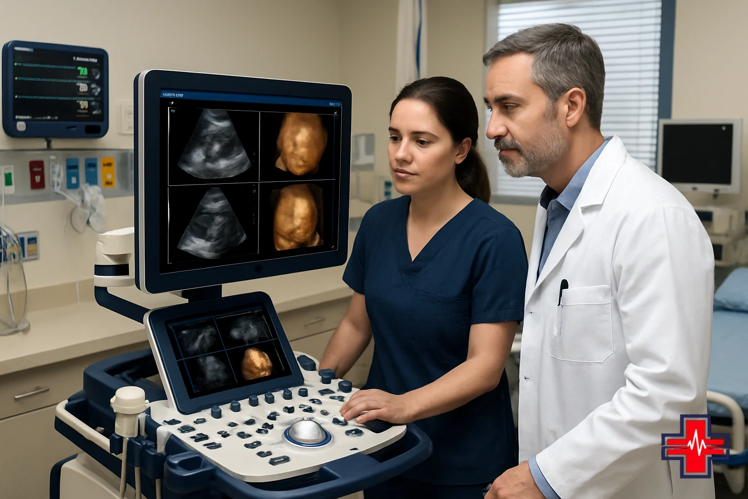
Image Acquisition and Display. The most fundamental difference is how images are created and presented. 2D ultrasound captures single planes in real time and displays them immediately as grayscale cross-sections. 3D ultrasound acquires a volume of data across multiple planes in rapid succession, then reconstructs that volume into a three-dimensional dataset that clinicians can rotate and slice digitally. This means a 2D exam is dynamic and responsive to the operator's real-time clinical questions, while a 3D exam involves a static acquired volume that is analyzed post-acquisition.
Visualization and Detail. 2D ultrasound excels at showing internal anatomy, blood flow (via Doppler), and functional movement of organs and the fetus. The grayscale format is familiar to clinicians and allows rapid, intuitive interpretation. 3D ultrasound, by contrast, excels at surface anatomy and spatial relationships. Surface renderings can reveal facial structure, limb position, and overall fetal morphology in ways that feel more intuitive to non-medical viewers. However, 3D imaging can obscure internal detail if deep tissue structures are the focus.
Examination Time and Workflow. A standard 2D prenatal ultrasound typically requires 15–30 minutes of active scanning, with the sonographer adjusting planes continuously to answer clinical questions in real time. A 3D volume acquisition adds time for the volumetric scan itself, plus additional time at the workstation for reconstruction and analysis. In busy clinical settings and emergency departments, the added time may not be practical or necessary.
Operator Skill and Equipment. Both modalities depend heavily on sonographer training and experience. A skilled 2D sonographer knows which planes to acquire and how to optimize image quality for diagnostic purposes. 3D sonography requires additional expertise in volume positioning, reconstruction algorithms, and post-processing software. Equipment quality also differs; high-end 3D systems capable of real-time volumetric acquisition are more expensive than conventional 2D machines.
Diagnostic Accuracy and Clinical Applications
The clinical value of any imaging modality ultimately rests on its ability to answer diagnostic questions accurately. When it comes to prenatal screening, 2D ultrasound has decades of evidence demonstrating its effectiveness in detecting fetal anomalies, assessing growth, evaluating placental position, and identifying pregnancies at risk. The detection rates for major congenital heart defects, neural tube defects, and other structural abnormalities are well established for 2D ultrasound when performed by trained operators using standardized protocols.
3D ultrasound can enhance diagnostic accuracy for specific anatomical questions—particularly regarding facial anatomy, complex cardiac defects, and precise volumetric measurements—but it does not universally improve detection of fetal anomalies over high-quality 2D imaging. Studies show that diagnostic accuracy depends much more on operator skill, equipment quality, clinical indication, and adherence to standardized scanning protocols than on simply choosing 3D over 2D. In other words, a poor-quality 3D exam may be less informative than an excellent 2D exam performed by an experienced sonographer.
For emergency scenarios—such as evaluating vaginal bleeding in pregnancy, assessing fetal heart rate following trauma, or confirming fetal viability in a threatened miscarriage—2D ultrasound with real-time Doppler assessment remains the standard. The ability to quickly acquire multiple planes, evaluate blood flow, and answer urgent clinical questions in minutes rather than requiring lengthy volumetric reconstruction makes 2D ultrasound the practical choice in time-critical situations. Emergency departments in Fort Worth, Keller, Alliance, and surrounding areas rely on 2D ultrasound capabilities to assess pregnant patients with acute symptoms.

Safety, Resolution, and Image Quality
Both 2D and 3D ultrasound are considered safe when performed according to established guidelines from the American Institute of Ultrasound in Medicine (AIUM) and the International Society of Ultrasound in Obstetrics and Gynecology (ISUOG). The safety of ultrasound in pregnancy is fundamentally about thermal and mechanical effects. Thermal index measures the potential for ultrasound energy to generate heat in tissue, while mechanical index reflects the potential for cavitation (bubble formation in tissue). Responsible clinical practice keeps both indices as low as reasonably achievable while obtaining diagnostic-quality images.
Image resolution refers to the smallest distance between two objects that can be distinguished as separate on an ultrasound image. Both 2D and 3D ultrasound are limited by the same physics: the wavelength of the sound used and the focusing properties of the transducer. In general, higher-frequency transducers provide better resolution but less penetration; lower-frequency transducers penetrate deeper but with lower resolution. A well-performed 2D ultrasound exam at 18–20 weeks of gestation, using appropriate transducer frequency and gain settings, achieves resolution sufficient to detect structures as small as 2–3 millimeters. 3D ultrasound does not inherently improve axial resolution; what it adds is the ability to view those structures from different angles and in surface detail.
Image quality in both modalities depends on maternal factors (body mass index, amniotic fluid volume), fetal position, transducer quality, system software, and sonographer technique. Artifacts—false images caused by reverberation, shadowing, or incomplete data—can occur in both 2D and 3D acquisitions and require experienced interpretation to avoid misdiagnosis. In emergency settings where rapid assessment is paramount, high-quality 2D imaging with real-time evaluation remains the most reliable and efficient approach.
When to Seek Emergency Ultrasound Care
Pregnant patients in Fort Worth and nearby communities—including Keller, Alliance, Saginaw, Haslet, Watauga, Roanoke, North Richland Hills, Haltom City, Bedford, Southlake, Grapevine, and Flower Mound—should seek urgent ultrasound evaluation for several conditions. Vaginal bleeding at any gestational age warrants immediate assessment to determine the location of bleeding, viability of the fetus, and presence of complications such as abruption or subchorionic hematoma. Severe abdominal or pelvic pain requires urgent imaging to rule out ectopic pregnancy, placental abruption, preterm labor, or other emergencies.
Absence of fetal movement or decreased fetal movement in the second and third trimester should prompt same-day evaluation, including ultrasound assessment of fetal heart rate, amniotic fluid volume, and placental function. Trauma or falls during pregnancy—even if the impact seems minor—warrant urgent ultrasound to assess for placental injury, abruption, or fetal distress. Signs of preterm labor, including regular uterine contractions, pelvic pressure, or vaginal fluid leakage before 37 weeks, require rapid imaging and fetal monitoring.
In these scenarios, ER Of Fort Worth provides 24/7 emergency ultrasound services using 2D imaging capability to rapidly answer critical questions about fetal viability, maternal safety, and need for higher-level care. The real-time nature and diagnostic power of 2D ultrasound make it the ideal modality for urgent assessment. Emergency room staff work quickly to stabilize the patient, perform targeted ultrasound imaging, and coordinate transfer to obstetric care or deliver results that guide outpatient follow-up.
If you are experiencing a life-threatening emergency, call 911 or go to the nearest emergency room immediately.
Frequently Asked Questions
Is 3D ultrasound necessary during pregnancy? No. For standard prenatal screening and monitoring, 2D ultrasound is the established clinical standard and is fully sufficient. 3D ultrasound may add value in specific clinical scenarios, such as evaluating suspected facial clefts or complex cardiac anatomy, but it is not routinely necessary and is not covered by insurance in most elective situations.
Can 3D ultrasound detect all fetal abnormalities that 2D can detect? Not necessarily. 3D ultrasound excels at surface anatomy but may not visualize internal structures as clearly as high-quality 2D imaging. Cardiac lesions, brain malformations, and renal abnormalities are often better assessed using 2D planes and Doppler evaluation. Diagnostic sensitivity depends more on operator skill and imaging protocol than on modality choice.
How is the Fort Worth ER equipped for obstetric ultrasound? ER Of Fort Worth maintains modern ultrasound equipment and trained sonographers capable of performing 2D ultrasound exams, including Doppler assessment of blood flow. This allows rapid evaluation of pregnant patients with acute symptoms and supports clinical decision-making regarding the need for higher-level obstetric care.
Is 3D ultrasound safe for the baby? Yes. When performed with appropriate attention to thermal and mechanical indices, 3D ultrasound is safe during pregnancy. However, routine use of 3D imaging simply for aesthetic purposes ("keepsake" imaging) is not recommended by major obstetric organizations, as there is no established medical benefit to offset the additional ultrasound exposure time.
Can I get a 3D ultrasound at an emergency room? Most emergency rooms, including ER Of Fort Worth, focus on 2D ultrasound for urgent assessment because it provides rapid answers to clinical questions. 3D imaging is typically available at specialized imaging centers or obstetric offices that have the equipment and time to perform elective volumetric studies.
Frequently Asked Questions
What is the main clinical advantage of 2D ultrasound over 3D in emergency pregnancy care?
2D ultrasound provides real-time, dynamic visualization of fetal heart rate, movement, and blood flow in minutes, making it ideal for rapid assessment of urgent obstetric concerns like bleeding, pain, or reduced fetal movement. 3D imaging requires additional time for volumetric acquisition and reconstruction, which is impractical in emergency settings.
Can 3D ultrasound replace 2D ultrasound for routine prenatal screening?
No. While 3D ultrasound can complement 2D imaging in certain cases, 2D ultrasound remains the clinical gold standard for routine prenatal screening because it has decades of evidence supporting its diagnostic accuracy, allows real-time assessment of fetal function, and is the basis for all established screening protocols.
Does 3D ultrasound provide better image resolution than 2D ultrasound?
Not inherently. Both modalities are limited by the same physics of sound wave propagation. 3D ultrasound adds the ability to view structures from multiple angles and create surface renderings, but axial resolution—the ability to distinguish closely-spaced structures—is determined by transducer frequency and does not differ between 2D and 3D systems.
Is it safe to choose 3D ultrasound instead of 2D for a keepsake image?
While 3D ultrasound is safe when performed with appropriate technique, major obstetric organizations do not recommend elective 3D imaging simply for aesthetic purposes, as there is no established medical benefit and the additional ultrasound exposure time is not justified. Routine 2D ultrasound is sufficient for standard prenatal care.
When would a doctor recommend 3D ultrasound as part of prenatal care?
3D ultrasound may be recommended when a specific anatomical question requires volumetric reconstruction and multi-planar viewing, such as suspected facial cleft, complex cardiac defect, or detailed fetal surface anatomy assessment. The decision is based on clinical indication and available equipment, not routine protocol.
What should I do if I have vaginal bleeding during pregnancy?
Seek urgent evaluation at a 24/7 emergency room like ER Of Fort Worth. Vaginal bleeding during pregnancy can indicate several conditions requiring immediate assessment, including threatened miscarriage, ectopic pregnancy, placental abnormalities, or abruption. Real-time ultrasound imaging is necessary to determine fetal viability and maternal stability.