
Benefits of 3D Ultrasound Imaging in Pregnancy
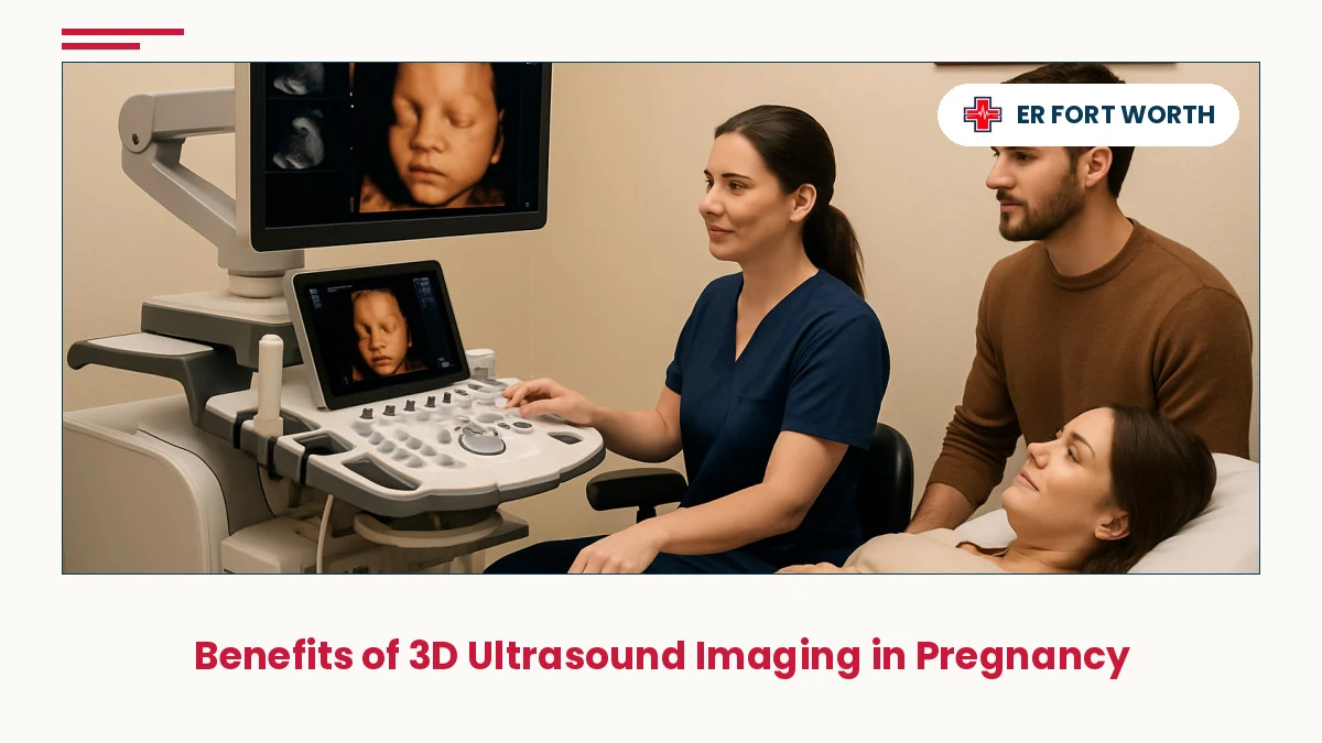
3D ultrasound imaging has transformed prenatal care by providing expectant parents with unprecedented clarity of fetal development and helping clinicians identify potential complications early. Learn how this advanced imaging technology enhances safety, bonding, and peace of mind during pregnancy.
In This Article
- What Is 3D Ultrasound Imaging?
- Fetal Bonding and Emotional Connection
- Early Anomaly Detection and Diagnostic Advantages
- Diagnostic Benefits for Pregnancy Monitoring
- Optimal Timing and Scheduling Recommendations
- When to Seek Emergency Ultrasound Imaging
- Safety Profile and Medical Evidence
- Frequently Asked Questions
⚡ Key Takeaways
- 3D ultrasound provides detailed facial and anatomical visualization that enhances emotional connection and parental bonding with the developing fetus.
- Advanced imaging helps clinicians detect structural anomalies and developmental variations earlier than traditional 2D ultrasound alone.
- Real-time 3D visualization improves diagnostic confidence and reduces the need for repeat imaging in many pregnancy evaluations.
- Expectant parents in Fort Worth and nearby communities can access 3D ultrasound services through specialized imaging centers and emergency facilities.
- While primarily diagnostic, 3D imaging creates memorable keepsakes that support healthy emotional attachment during pregnancy.
What Is 3D Ultrasound Imaging?
Three-dimensional ultrasound represents an evolution in prenatal imaging technology that builds upon conventional two-dimensional scanning. While traditional 2D ultrasound displays a flat, cross-sectional slice of the fetus, 3D ultrasound captures volumetric data—entire blocks of tissue information—that can be reconstructed and rotated to show multiple planes and perspectives simultaneously.
The technology works by rapidly acquiring hundreds of 2D images from different angles within a specific region, then using advanced software to compile these datasets into realistic, three-dimensional representations. This approach allows sonographers and physicians to examine fetal anatomy from virtually any angle, rotate the image in real time, and measure structures with exceptional precision. Modern 3D systems often integrate 4D capability—adding the dimension of motion—so parents and clinicians can observe the fetus moving, breathing, and responding in near real-time.
For expectant families in Fort Worth, Keller, Alliance, Saginaw, Haslet, and surrounding North Texas communities, access to 3D ultrasound services has expanded significantly. Fort Worth Emergency Room and other specialized imaging centers now routinely offer 3D imaging as part of comprehensive prenatal assessment.
Fetal Bonding and Emotional Connection
One of the most profound benefits of 3D ultrasound imaging in pregnancy is the opportunity it creates for enhanced parental bonding with the developing fetus. Unlike 2D images, which require interpretation and visualization skills to understand, 3D images reveal recognizable facial features, hand positions, and body posture in ways that feel immediately familiar and emotionally resonant.
Seeing a clear, lifelike image of the baby's face—with visible features like nose, lips, and eyes—transforms the pregnancy from an abstract medical concept into a tangible, relational reality. Parents report that viewing 3D ultrasound images strengthens their emotional attachment and sense of connection to the pregnancy. This phenomenon has been observed across cultures and socioeconomic backgrounds, suggesting a universal human response to recognizing facial features and body form.
For expectant mothers and families who may experience anxiety, grief, or complicated feelings during pregnancy, the opportunity to see and bond with the fetus through 3D imaging can provide reassurance and psychological support. The keepsake images and videos that result from 3D ultrasound sessions often become treasured mementos that families preserve throughout the child's life.
Research in maternal-fetal psychology indicates that positive bonding experiences during pregnancy correlate with healthier postpartum adjustment and early parent-child attachment patterns. While 3D ultrasound alone is not a therapeutic intervention, the bonding opportunity it facilitates can support overall maternal psychological health.
Early Anomaly Detection and Diagnostic Advantages
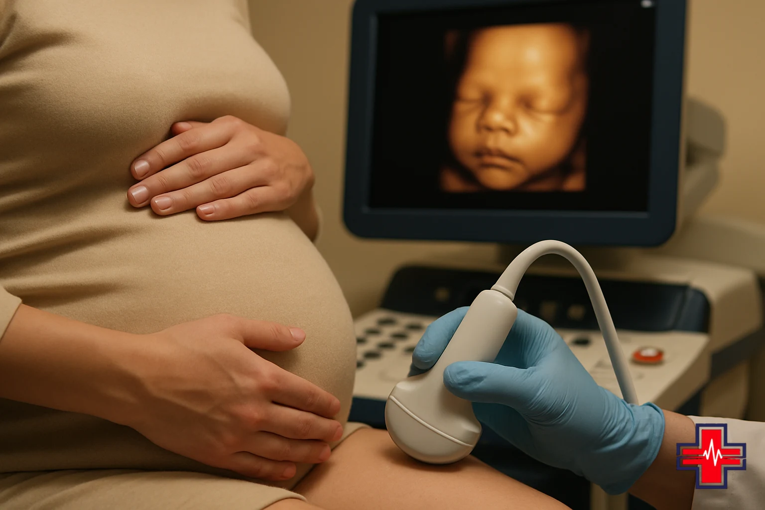
From a clinical perspective, 3D ultrasound imaging provides significant diagnostic advantages for detecting fetal anomalies and structural variations. The ability to examine anatomy from multiple planes simultaneously allows sonographers to identify subtle abnormalities that might be missed on 2D imaging alone.
Certain fetal structures benefit particularly from 3D visualization. The fetal face and palate can be assessed for cleft palate or other oro-facial anomalies with greater sensitivity. The spine can be evaluated in three dimensions to detect segmentation abnormalities, asymmetries, or neural tube defects. Cardiac anatomy becomes clearer when viewed in three-dimensional reconstructions, improving the detection of congenital heart malformations. Limb and hand anatomy can be thoroughly examined to assess for polydactyly (extra fingers or toes), syndactyly (fused digits), or other skeletal variations.
The diagnostic benefits extend beyond structural anomaly detection. 3D imaging also supports more accurate biometric measurements—precise estimation of fetal weight, head circumference, and organ size. These measurements are crucial for identifying growth restriction, fluid imbalances, and developmental delays. The volumetric data can be stored and reviewed later, allowing multiple physicians to contribute to diagnostic interpretation without requiring the patient to return for repeat imaging.
For families navigating complex pregnancies or elevated-risk situations, the diagnostic confidence provided by 3D ultrasound can reduce anxiety by clarifying exactly what clinicians can see—and reassuring families when careful examination reveals normal development.
Diagnostic Benefits for Pregnancy Monitoring
Beyond bonding and anomaly detection, 3D ultrasound offers several additional diagnostic benefits that enhance comprehensive pregnancy care:
- Improved Placental Assessment: The placenta's position, thickness, vascularity, and attachment can be evaluated three-dimensionally, helping clinicians identify potential complications like placenta previa or placental insufficiency earlier.
- Enhanced Amniotic Fluid Evaluation: 3D imaging allows more precise measurement of amniotic fluid volume and distribution, critical for identifying polyhydramnios (excess fluid) or oligohydramnios (insufficient fluid) that may signal underlying concerns.
- Detailed Organ Visualization: The fetal kidneys, bladder, lungs, liver, and other organs can be examined in three dimensions, enabling detection of cystic structures, enlargement, or absence that warrants intervention or monitoring.
- Reduced Need for Repeat Scans: Because 3D data can be reconstructed and reexamined from any angle, many diagnostic questions can be answered from a single imaging session, reducing radiation exposure and appointment burden.
- Superior Communication with Patients: Showing parents a realistic 3D image of their baby fosters understanding of prenatal findings and improves informed decision-making regarding management options.
For expectant parents in North Richland Hills, Roanoke, Watauga, Haltom City, Bedford, Southlake, Grapevine, and Flower Mound, access to facilities offering comprehensive 3D prenatal imaging ensures they receive state-of-the-art diagnostic support throughout pregnancy.

Optimal Timing and Scheduling Recommendations
The timing of 3D ultrasound imaging significantly influences image quality and diagnostic yield. Expectant parents often ask when the optimal window exists for 3D ultrasound benefits for pregnancy assessment.
First Trimester (Weeks 11–14): Early 3D imaging can assess nuchal translucency (the fluid pocket at the back of the fetal neck), confirm dating, detect multiple pregnancies, and verify fetal cardiac activity. However, image quality is typically lower than in later trimesters due to smaller fetal size and more variable maternal tissue quality.
Second Trimester (Weeks 18–24): This represents the golden window for comprehensive 3D ultrasound. The fetus is large enough to visualize detailed anatomy, facial features are well-formed and recognizable, and most major organs have sufficiently developed. The detailed anomaly scan—often called the anatomy scan—is routinely performed during this period, and 3D imaging enhances the diagnostic value of this critical screening.
Third Trimester (Weeks 28–36): Later 3D imaging can assess fetal growth, amniotic fluid volume, placental position, and prepare for delivery planning. However, image quality may decrease slightly due to increased fetal size and reduced amniotic fluid volume relative to fetal volume.
Most prenatal imaging programs recommend elective 3D ultrasound between weeks 20–24 for optimal balance of anatomical detail, image quality, and parental bonding opportunity. However, clinical indications—such as suspected anomalies, growth concerns, or complications—may warrant 3D imaging at any stage of pregnancy.
When to Seek Emergency Ultrasound Imaging
If you are experiencing a life-threatening emergency, call 911 or go to the nearest emergency room immediately.
While many 3D ultrasound procedures are scheduled electively, certain pregnancy complications warrant urgent or emergency imaging evaluation. Expectant parents in Fort Worth and surrounding areas should seek immediate emergency care if experiencing:
- Vaginal bleeding of any amount after the first trimester, which may indicate placental abruption, preterm labor, or other serious complications
- Severe abdominal or pelvic pain that does not resolve with rest or standard pain management
- Sudden gush of fluid suggestive of amniotic membrane rupture
- Absence of fetal movement after quickening has been established (typically after week 20)
- Severe headache with vision changes or elevated blood pressure concerning for preeclampsia
- Signs of infection including fever, chills, or malaise combined with pregnancy symptoms
Fever treatment ER services at ER Of Fort Worth include obstetric triage and imaging for pregnant patients. Online check-in is available to expedite your arrival if you are able to safely travel to the facility. The 24/7 emergency room at Heritage Trace Parkway can assess these urgent concerns rapidly and coordinate appropriate imaging and care.
Safety Profile and Medical Evidence
Expectant parents frequently ask whether 3D ultrasound imaging is safe during pregnancy—a question grounded in appropriate caution about fetal well-being. The medical evidence supporting the safety of diagnostic ultrasound, including 3D applications, is robust.
Unlike ionizing radiation (X-ray, CT scan), diagnostic ultrasound uses sound waves and does not expose the fetus to radiation. The ultrasound equipment emits energy at power levels that are deliberately kept below thresholds known to cause thermal or mechanical injury to tissue. Extensive research spanning decades has not identified harmful effects from diagnostic ultrasound exposure in utero at clinically used power settings.
Major medical organizations, including the American College of Obstetricians and Gynecologists (ACOG), the CDC, and the American Institute of Ultrasound in Medicine (AIUM), affirm that diagnostic ultrasound is safe for prenatal evaluation when performed by trained sonographers using appropriate equipment and protocols. The key safety principle is using ultrasound for medical indication—not for frivolous or excessive recreational imaging without clinical purpose.
3D ultrasound technology does not expose the fetus to additional radiation or substantially greater energy than 2D ultrasound. The volumetric reconstruction that creates the 3D image occurs within the ultrasound system itself; no additional energy is applied to fetal tissue. For expectant families seeking 3D imaging for bonding purposes combined with diagnostic evaluation, the risk-benefit analysis clearly favors the imaging when performed at established facilities with qualified personnel.
Expectant parents interested in learning more about ultrasound safety can review resources from MedlinePlus, which provides evidence-based information about prenatal imaging procedures and their safety profiles.
Frequently Asked Questions
What is the main difference between 3D and 4D ultrasound?
3D ultrasound captures volumetric anatomical data that can be rotated and viewed from multiple angles, while 4D adds real-time motion, allowing you to see the baby moving and breathing as the image is being scanned. Both use the same basic technology; 4D simply includes the element of motion over time.
Can 3D ultrasound detect all birth defects?
While 3D ultrasound improves detection of many structural anomalies—particularly those involving facial features, limbs, and organ shape—some conditions cannot be visualized prenatally or may not manifest until later in development. Comprehensive prenatal screening combines 3D imaging with other tests like maternal serum screening and, when indicated, genetic counseling.
Is 3D ultrasound covered by insurance?
Insurance coverage varies by plan and whether the imaging is performed for medical indication versus elective bonding purposes. Medically indicated 3D ultrasound—ordered by a physician for diagnostic reasons—is more likely to be covered. Elective bonding or keepsake imaging is often out-of-pocket. Contact your insurance provider and imaging facility to clarify coverage before scheduling.
How long does a 3D ultrasound appointment typically take?
A comprehensive 3D ultrasound session usually takes 30–45 minutes, depending on fetal position, imaging goals, and whether both diagnostic assessment and bonding imaging are included. Appointments may run longer if detailed evaluation of specific concerns is required or if the fetus is in a position that limits visualization.
Can I bring family members to my 3D ultrasound appointment?
Most facilities welcome one or two support persons to share the bonding experience. Some imaging centers have specific policies regarding additional guests, particularly in clinical settings like emergency rooms. Check with your facility beforehand regarding their visitor and accompaniment policies.
Should I schedule 3D ultrasound if my pregnancy is high-risk?
High-risk pregnancies often benefit from the enhanced diagnostic information provided by 3D ultrasound. Discuss with your obstetrician whether 3D imaging would be beneficial for your specific situation. Many high-risk pregnancies warrant specialized imaging to support careful monitoring and management planning.